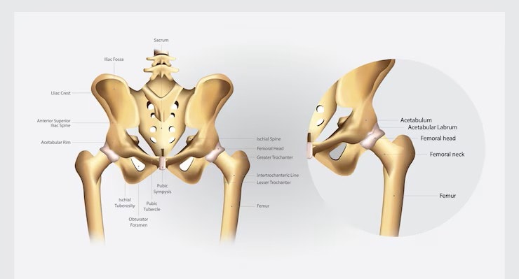
Hip bone anatomy involves understanding the three main components: the ilium, ischium, and pubis. These bones form a structure crucial for movement, stability, and weight-bearing. This article will explore these components, their articulations, blood supply, innervation, muscle attachments, and developmental aspects.
Key Takeaways
The hip bone comprises three main parts: ilium, ischium, and pubis, working together to support the upper body and facilitate lower limb movement.
Key joints like the hip joint, sacroiliac joint, and pubic symphysis enhance the stability and flexibility of the pelvis, crucial for various movements and activities.
Understanding hip bone anatomy is essential in clinical contexts, including diagnosing fractures, performing hip replacements, and managing trauma patients.
The Hip Bone Structure
The hip bone, also known as the pelvic girdle, is a complex structure formed by the fusion of three bones: the ilium, ischium, and pubis, collectively referred to as the os coxae. These pelvic bones develop through a fusion process that begins in the embryonic stage and continues into adolescence, resulting in the mature hip bone that articulates with the axial skeleton.
The hip bones play a crucial role in forming the pelvis, providing support for the upper body and facilitating movement of the lower limbs. Each component of the hip bone has unique features and functions that contribute to the overall stability and mobility of the human pelvis.
Hip Bone Diagram

Ilium
The ilium is the largest part of the hip bone, characterized by its blade-like shape. It forms the upper portion of the acetabulum, where the femur articulates. Key landmarks of the ilium include the anterior superior iliac spine (ASIS) and the posterior superior iliac spine (PSIS), which mark the front and back endpoints of the iliac crest.
The iliac crest itself is the upper boundary of the ilium, serving as an attachment point for various muscles and ligaments, such as the gluteus maximus, gluteus medius, and gluteus minimus.
Ischium
The ischium is another key component of the hip bone anatomy, situated below the ilium and contributing to the lower and back part of the pelvis. One of its most notable features is the ischial tuberosity, which acts as a support point when sitting.
Additionally, the ischial spine serves as an attachment point for ligaments, while the lesser sciatic notch allows for the passage of nerves and vessels. These features highlight the ischium’s importance in weight-bearing and providing attachment sites for muscles and ligaments.
Pubis
The pubis is the front part of the hip bone. It is made up of a body and two rami. The superior pubic ramus connects to the ilium, forming part of the acetabulum, while the inferior pubic ramus connects to the ischium. The pubic body forms the central part of the pubis and the anterior boundary of the pelvic girdle.
The pubic symphysis, located at the front of the pelvis, is a cartilaginous joint that allows for slight movement, enhancing flexibility and accommodating activities like walking and childbirth.
Articulations and Joints
The hip bone’s connections with the rest of the skeletal system are vital for its function. These articulations include the hip joint, sacroiliac joint, and pubic symphysis, each playing a crucial role in the anatomy of the hip. These joints contribute to the overall stability and flexibility of the pelvis, enabling a wide range of movements and weight-bearing activities.
Hip Joint
The hip joint, classified as a ball-and-socket joint, is where the hip bone articulates with the femur. This joint allows for a diverse range of motion, essential for activities such as walking, running, and jumping. The femoral head fits into the acetabulum of the hip bone, forming a stable yet flexible connection.
This joint’s unique structure enables it to bear the body’s weight while providing extensive mobility in multiple directions.
Sacroiliac Joint
The sacroiliac joint connects the sacrum to the ilium, providing stability and strength to the pelvis. This joint plays a vital role in transferring weight from the upper body to the lower limbs, making it essential for maintaining balance and stability during movement.
The sacroiliac joint’s robust structure ensures that it can withstand significant forces while allowing slight movements to accommodate various activities.
Pubic Symphysis
The pubic symphysis is a cartilaginous joint where the left and right pubic bones meet, allowing for slight movement and flexibility. This joint is particularly important during activities such as walking and childbirth, where slight adjustments in the pelvis are necessary. The fibrocartilage at the pubic symphysis provides a cushion that absorbs shock and maintains stability.
This joint ensures that the pelvis can accommodate changes in position and pressure without compromising its structural integrity.
Blood Supply and Innervation
The vascular and nervous systems surrounding the hip bone are crucial for its function and health. These systems ensure that the hip region receives adequate blood supply and nerve signals, which are essential for maintaining the joint’s health and facilitating movement.
Femoral Artery
The femoral artery is the principal vessel delivering oxygenated blood to the lower limbs, originating near the groin and extending down to the knee. This artery is a primary source of blood flow to the hip region, supplying essential nutrients and oxygen to the hip joint and surrounding structures.
Branches of the femoral artery, such as the medial and lateral circumflex arteries, play significant roles in ensuring the hip’s adequate blood supply.
Obturator Nerve
The obturator nerve is crucial for motor control and sensory perception in the medial thigh. Originating from the lumbar plexus, specifically from the L2, L3, and L4 spinal nerves, it travels through the pelvis to exit via the obturator foramen.
This nerve innervates the adductor muscles of the thigh, facilitating actions like hip adduction and playing a significant role in stabilizing the pelvis during movement.
Sciatic Nerve
The sciatic nerve, the largest nerve in the body, extends from the lower back into the lower limb, influencing both motor function and sensation in the posterior thigh and leg. It plays a crucial role in relaying signals between the hip and lower limb, impacting movement and sensory perception.
Any injury or compression of the sciatic nerve can significantly affect mobility and cause severe pain, highlighting its importance in the overall function of the lower body.
Muscular Attachments
The hip bone serves as an anchor point for various muscle groups that facilitate movement and stability. These muscles are essential for performing daily activities and maintaining balance, underscoring the importance of the hip bone in overall mobility.
Gluteal Muscles
The gluteal muscles, including the gluteus maximus, medius, and minimus, originate from the posterior surface of the ilium and play vital roles in movement and stability. The gluteus maximus is the primary extensor of the thigh, while the gluteus medius stabilizes the pelvis and inserts at the greater trochanter.
The tensor fascia lata, originating from the anterior iliac crest, assists in abducting and medially rotating the lower limb. These muscles are crucial for activities such as walking, running, and maintaining an upright posture.
Adductor Muscles
The adductor muscles primarily function to adduct the thigh, playing a critical role in stabilizing the pelvis during movement. These muscles arise from the pubis and ischium, attaching mainly to the femur’s linea aspera. The obturator nerve innervates these muscles, facilitating hip movement and providing sensory input from the inner thigh region.
Key adductors include the adductor longus and adductor magnus, which are essential for activities that require thigh movement toward the body’s midline.
Pelvic Floor Muscles
Pelvic floor muscles play a crucial role in supporting pelvic organs and maintaining core stability. These muscles are directly connected to the hip bone through various muscle attachments, influencing movement and stability.
Strong pelvic floor muscles contribute to:
Improved bladder control
Improved bowel control
Enhanced sexual function
Overall health
Maintaining these muscles’ health through exercises can prevent dysfunction and enhance overall pelvic health.
Development and Variants
The development of the hip bone begins in the embryonic stage and transitions through various phases until reaching maturity during puberty. Understanding these developmental stages and the common anatomical variants is crucial for clinical practice and diagnosing conditions related to the hip bone.
Embryological Development
The hip bone begins its development as a mesodermal condensation in the lower limb bud during early fetal stages. Initially, the ilium, ischium, and pubis are separate structures. As the fetus grows, these bones undergo significant changes, gradually fusing to form the acetabulum by puberty.
This process ensures the structural integrity and functionality of the hip bone, providing a stable base for the lower limbs.
Sexual Dimorphism
Sexual dimorphism in hip bones illustrates distinct anatomical features between males and females. Male hip bones are generally heavier and thicker, adapted for bipedal locomotion. In contrast, female hip bones are lighter and designed to facilitate childbirth.
The pelvic inlet in females is wider and oval-shaped, while in males, it is narrower and heart-shaped. Additionally, the subpubic angle is broader in females, and the greater sciatic notch is wider to aid in childbirth.
Common Variants
Common pelvic shapes, including gynaecoid, android, platypelloid, and anthropoid, each have specific characteristics and clinical implications. The gynaecoid pelvis, the most common type in females, is characterized by a wide transverse diameter, facilitating easier childbirth.
The android pelvis, heart-shaped, is predominantly seen in males and is narrower, affecting childbirth mechanics. Platypelloid and anthropoid pelvis shapes also present unique challenges and advantages in obstetrics.
Clinical Relevance
Understanding the clinical relevance of hip bone anatomy is paramount for diagnosing and treating various conditions. From pelvic fractures to hip replacement surgeries, the hip bone’s structure and function are critical in medical practice.
These treatments and conditions highlight the importance of a thorough understanding of hip bone anatomy.
Pelvic Fractures
Pelvic fractures typically result from high-impact trauma and can lead to a range of complications, necessitating thorough assessment and prompt management. Common types include stable and unstable fractures, with unstable fractures posing a higher risk for internal injuries and often requiring surgical intervention to prevent complications such as hemorrhage or organ damage.
These fractures highlight the importance of the pelvis in protecting internal organs and supporting the body’s weight.
Hip Replacement
Hip replacement surgery is often performed to relieve pain and restore function in individuals with severe hip joint damage. This procedure involves replacing the damaged hip joint with artificial components made from metal, ceramic, or plastic.
Total hip arthroplasty, a common form of hip replacement, has been shown to improve pain management and functional outcomes significantly. Early surgical intervention is linked to better recovery rates and lower complication risks.
Major Trauma Patients
Major trauma patients with hip injuries often require a multidisciplinary approach for effective management and rehabilitation. Compromised pelvic stability can lead to complications in trauma management, influencing treatment decisions and outcomes.
An improved understanding of pelvic anatomy and stability can enhance protocols in trauma care related to hip injuries, ensuring better patient outcomes.

