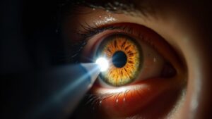
Pupil response offers a window into brain health, revealing concealed clues about injury severity. Whenever light hits the eyes, the brain signals pupils to constrict—a reflex that weakens if trauma disrupts neural pathways.
Uneven pupil size or sluggish reactions often signal swelling, bleeding, or pressure buildup. In contrast to subjective checks, automated pupillometry detects subtle changes missed by the naked eye, assisting doctors spot trouble sooner. These tiny black circles hold answers that could save lives, but what occurs when they cease responding normally?
The Science Behind Pupil Response and Brain Function
Because the eyes provide a direct window into brain function, perception of pupil response aids in detecting even subtle neurological disruptions. The pupillary light reflex, governed by the brain’s autonomic nervous system, reveals how well the brain processes signals.
In patients with traumatic brain injury (TBI), even mild concussions can disrupt this reflex, causing uneven pupil size or delayed reactions to light. The Glasgow Coma Scale often includes pupillary checks because changes gesture at deeper brain dysfunction.
Automated tools now measure precise pupil movements, capturing issues traditional exams could miss. Since the brain’s midbrain regulates pupil control, damage there can weaken responses before other symptoms manifest. This makes pupillary tests crucial for spotting concealed injuries, offering clues when speech or movement exams fall short.
How Pupil Testing Complements Traditional Brain Injury Assessments
Pupil testing acts like an X-ray for the brain, uncovering obscured injuries that standard exams could overlook. Traditional assessments, like symptom reports or balance tests, often miss subtle but critical signs of traumatic injury. Automated pupillometry measures the pupillary light reflex (PLR) with precision, detecting irregularities that hint at increased intracranial pressure or autonomic dysfunction.
- Objective Insights: Unlike subjective symptom reports, pupil response provides measurable data, reducing bias in diagnosis.
- Early Detection: Slower constriction or delayed expansion can signal concussion before other symptoms arise.
- Monitoring Tool: Repeated testing tracks recovery, helping clinicians decide the appropriate time to return to activities.
Key Pupillary Metrics Used in Clinical Evaluations
Pupil size assessment provides critical information about autonomic nervous system function, with abnormal measurements often signaling brain injury. Light reflex evaluation scrutinizes how quickly pupils constrict when exposed to light, where delayed responses can indicate neurological impairment.
These metrics offer objective data that helps clinicians detect subtle changes in brain function that manual exams could miss.
Pupil Size Assessment
How crucial is pupil size when evaluating someone with a potential brain injury? Pupil size is a critical metric, offering immediate clues about neurological function. Abnormalities in pupil size or reactivity can signal serious issues like brainstem damage or increased intracranial pressure.
Normal Range: Healthy pupils typically measure 2–8 mm, adjusting to light (pupillary constriction) or darkness.
Warning Signs: Unequal sizes (anisocoria) or nonreacting pupils may indicate trauma or nerve damage.
Autonomic Clues: Pupil size reflects autonomic nervous system activity—dilated or constricted pupils without cause suggest dysfunction.
Pupillary abnormalities, like fixed dilation, often precede severe consequences, making timely assessment essential. Subtle changes in pupil reactivity can differentiate minor injuries from life-threatening conditions. Clinicians rely on these metrics to guide urgent care decisions, emphasizing their role in brain injury evaluations.
Light Reflex Evaluation
As time passes, doctors scrutinize how the pupils respond to light—a speedy test that divulges concealed issues, should someone’s brain potentially be harmed. The light reflex assessment measures pupil response, including size changes and speed, to detect problems like head injury or coma.
Sluggish or uneven reactions may indicate brain damage, affecting the Glasgow Outcome Scale score. Tools like quantitative pupillometry track pupillary dilation exactly, offering clearer data than manual checks. This helps anticipate recovery, especially when paired with the Coma Scale.
Quick, non-invasive, and revealing, this test gives critical clues about neurological health. Even subtle changes matter, guiding treatment decisions and monitoring progress after trauma. Comprehension of these responses can mean better care and improved outcomes for patients.
Pupil Abnormalities and Their Prognostic Value in TBI
- Fixed and Dilated Pupils: A sign of increased intracranial pressure, often linked to poor recovery.
- Unequal Pupil Size (Anisocoria): Suggests brain stem compression, requiring immediate intervention.
- Sluggish Light Reflex: Slower responses hint at mild to moderate damage, guiding treatment urgency.
These abnormalities help doctors predict recovery, offering families clarity during uncertain times. While alarming, prompt detection improves care, turning a small observation into a life-saving clue.
Monitoring pupils remains a simple yet powerful tool in TBI management.
Comparing Objective Pupillometry With Subjective Neurological Exams
Objective pupillometry and traditional neurological exams offer different ways to measure pupil response in brain injury cases. Objective pupillometry utilizes precise devices to track pupil size and reactivity, eliminating human error from brain injury assessments.
Subjective neurological exams rely on a clinician’s observation during a neurological examination, which can vary based on experience or lighting conditions. While clinical evaluation remains standard, objective pupillometry provides consistent, numerical data on pupil response, helping detect subtle changes missed by manual checks.
Both methods aim to identify brain dysfunction, but the automated approach reduces guesswork. Combining them improves accuracy, ensuring better monitoring in critical cases. This dual approach helps tailor treatment, offering clearer insights into a patient’s condition with fewer uncertainties.
Future Applications of Pupil Analysis in Brain Injury Care
Though pupillometry is already helping doctors assess brain injuries, its future uses could transform how care is delivered.
- Machine learning could refine pupil response as a reliable indicator for acute brain injuries, utilizing logistic regression to model risk factors faster than manual analysis.
- Automated tracking of cranial nerve function through pupillometry could detect subtle damage missed by traditional exams, especially in concussion cases.
- Expanding demographic databases will enable doctors to compare individual results against broader norms, boosting accuracy in brain injury diagnosis.
Researchers have used logistic regression to link pupil metrics like constriction speed with neurological outcomes. Multi-modal testing combining pupillometry with other tools may soon offer clearer insights into brain dysfunction, helping tailor recovery plans. Standardized protocols will ensure consistent measurements across clinics, making pupillometry a mainstream diagnostic tool.
Conclusion
The article should flow seamlessly.
Pupil response reveals what the naked eye often misses—tiny changes hinting at serious brain trouble. Could those fleeting milliseconds of sluggish reaction time signal swelling or pressure before other symptoms arrive? Automated measurements now spot dangers human exams may overlook, offering families clearer answers during alarming moments. While not a standalone solution, this tool adds critical data to the puzzle, ensuring subtle red flags don’t slip through the cracks as every instant matters.


